
Quantitative Neuromuscular Monitoring: The Essential Guide
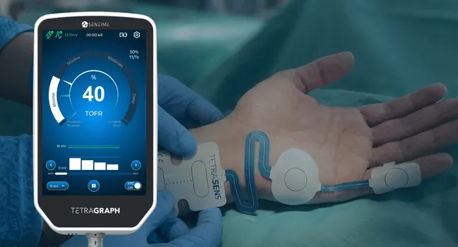
Residual neuromuscular block remains one of the most common and preventable patient safety risks in anesthesia. The challenge is that clinicians cannot reliably detect residual paralysis through observation alone.
The critical issue is the “zone of blind paralysis”. Between a train-of-four (TOF) ratio of 0.4 and 0.9, patients may appear clinically recovered while still experiencing meaningful neuromuscular impairment. Head-lift tests, grip-strength assessments, and visual evaluation of fade cannot detect this level of residual block. Only quantitative monitoring can provide the objective data needed to confirm recovery. [1,2]
This reality drove the 2023 American Society of Anesthesiologists (ASA) recommendations, which strongly support quantitative neuromuscular monitoring whenever nondepolarizing neuromuscular blocking agents are used. [1]
The Problem with Qualitative Monitoring and Why Guidelines Changed
Historically, anesthesia providers relied on qualitative monitoring techniques, including tactile or visual assessment of TOF fade, head-lift tests, grip strength, and clinical observation.
These methods have significant limitations. Research has shown that qualitative assessment becomes unreliable once TOF ratios exceed approximately 0.4. In fact, many patients can successfully perform a 5-second head lift despite clinically significant residual block. One study found that up to 80% of patients with TOF ratios below 0.7 could still complete the test [2].
The result is postoperative residual curarization (PORC), which remains surprisingly common. Without quantitative monitoring, studies report that 30% to 65% of patients arrive in the post-anesthesia care unit (PACU) with residual neuromuscular block. [3]
This is why ASA guidance shifted toward objective measurement. Latest quantitative monitoring technologies do not estimate recovery. It provides a numerical value clinicians can use to make informed reversal and extubation decisions.
What Quantitative Monitoring Actually Measures
Quantitative neuromuscular monitoring measures the train-of-four ratio, the accepted standard for assessing recovery from neuromuscular block.
The monitor delivers four supramaximal electrical pulses. When all four twitches are present, the TOF ratio is calculating by comparing the fourth twitch (T4) with the response to the first twitch (T1). The resulting TOF ratio is expressed as a value between 0 and 1.0. A TOF ratio of at least 0.9 is considered necessary to confirm adequate recovery before extubation.

Several technologies can generate TOF measurements:
- Electromyography (EMG) measures the compound muscle action potential generated by the electrical response of muscle to nerve stimulation and does not depend on thumb movement.
- Acceleromyography (AMG) measures acceleration of the thumb but requires unrestricted movement and often requires normalization because TOF values can exceed 1.0.
- Mechanomyography (MMG) measures force generation and remains the traditional research gold standard, but it is not practical for routine operating room use.
Many institutions are adopting EMG-based monitoring because it provides objective measurements without requiring unrestricted thumb movement.
The 2023 ASA Guidelines: 8 Key Recommendations
The 2023 ASA recommendations establish the current clinical standard for neuromuscular monitoring and recovery assessment [1].
- Do not rely solely on clinical signs or physical tests to assess recovery.
- Use quantitative monitoring rather than qualitative monitoring whenever possible.
- Confirm a TOF ratio of at least 0.9 before extubation.
- Monitor at the adductor pollicis muscle for the most clinically relevant assessment.
- Avoid relying on eye-muscle monitoring to determine recovery from block.
- Consider sugammadex over neostigmine for reversal of deep, moderate, or shallow block induced by rocuronium or vecuronium.
- Neostigmine is a reasonable alternative to sugammadex at minimal block.
- Atracurium/cisatracurium: reverse with neostigmine and confirm TOF ≥0.9 before extubation.
Importantly, quantitative monitoring not only identifies patients who need reversal, it also identifies patients who do not. One study found that 40% of monitored patients achieved spontaneous recovery to TOF ≥ 0.9 and did not require sugammadex [4]. Those drug cost savings are only possible when objective data are available.
EMG Technology: Why It's the Right Choice for Modern ORs
As hospitals seek to align with ASA recommendations, many are selecting EMG-based monitoring because it fits both clinical and operational needs.
EMG offers several advantages:
- Measures the evoked compound muscle action potential (CMAP)
- Does not require a freely moving thumb
- Works in robotic, laparoscopic, prone, and tucked-arm procedures
- Provides objective measurements
- Supports efficient OR workflows
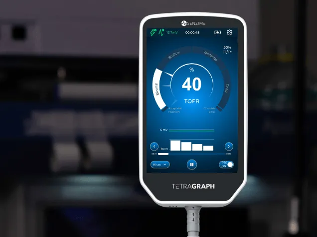
TetraGraph® is a purpose-built EMG monitoring system designed for routine clinical use. The FDA-cleared device uses a compact electrode design and has demonstrated 97.9% agreement with MMG benchmark measurements. [5]
For organizations focused on documentation and compliance, TetraGraph offers several integrations enabling automatic transfer of monitoring data into the electronic medical record. This can help to reduce manual charting requirements while improving visibility into monitoring practices.
Most hospitals can implement quantitative monitoring with minimal workflow disruption.
A typical approach includes:
- Evaluating available monitoring technologies, with EMG offering the greatest flexibility across surgical settings.
- Comparing sensor costs against potential savings from optimized sugammadex utilization and reduced complications.
- Training staff in electrode placement and device operation.
- Integrating monitoring data into EMR systems
- Tracking TOF documentation rates and postoperative outcomes as quality metrics.
The impact can be substantial. In one large multi-campus quality improvement initiative, TOF documentation rates increased from 42% to 83%, while postoperative respiratory complications fell by 41% [6].
Quantitative neuromuscular monitoring is no longer simply a technology upgrade. It is the most reliable way to detect residual block, support ASA guideline compliance, improve patient safety, and optimize perioperative care.
Learn how EMG-based monitoring can support safer recoveries, stronger documentation, and easier ASA guideline compliance.
Please note: This page is intended for healthcare professionals. It is primarily tailored to the U.S. market, but may also be relevant for other regions.
References
- Thilen SR, Weigel WA, Todd MM, et al. 2023 American Society of Anesthesiologists practice guidelines for monitoring and antagonism of neuromuscular blockade: a report by the American Society of Anesthesiologists Task Force on Neuromuscular Blockade. Anesthesiology. 2023;138(1):13-41. doi: 10.1097/ALN.0000000000004379 .
- Kopman AF, Yee PS, Neuman GG. Relationship of the train-of-four fade ratio to clinical signs and symptoms of residual paralysis in awake volunteers. Anesthesiology. 1997;86(4):765-771. doi:10.1097/00000542-199704000-00004.
- Murphy GS, Brull SJ. Residual neuromuscular block: lessons unlearned. Part I: definitions, incidence, and adverse physiologic effects of residual neuromuscular block. Anesth Analg. 2010;111(1):120-128.
- Haberkorn S, Twite M, Klockau K, Whitney G, Faulk DJ. Quantitative monitoring maximizes cost-saving strategies when antagonizing neuromuscular block with sugammadex. Cureus 2024; 16(9):e68551.
- Ebert TJ, Vogt JA, Kaur R, et al. Train-of-four ratio, counts and post tetanic counts with the Tetragraph electromyograph in comparison to mechanomyography. J Clin Monit Comput. 2025;39(1):149–156. doi:10.1007/s10877-024-01225-3.
- Goriacko P, Chao J, Fassbender P, Rudolph M, Beechner P, Shukla H, Yaghdjian V, Choice C, Aroh F, Sinnett M, Karaye I, Eikermann M. Optimizing neuromuscular block monitoring and reversal: A large-scale quality improvement initiative in a diverse healthcare setting, Journal of Clinical Anesthesia, Volume 101, 2025, 111709, ISSN 0952-8180, https://doi.org/10.1016/j.jclinane.2024.111709 .