Why EMG Signal Strength Matters and How TetraGraph® is Superior in Assessing Reliable Recovery
September 24, 2026
Blog by Linnea Troeng, Sr Global Manager, Products and Business Development
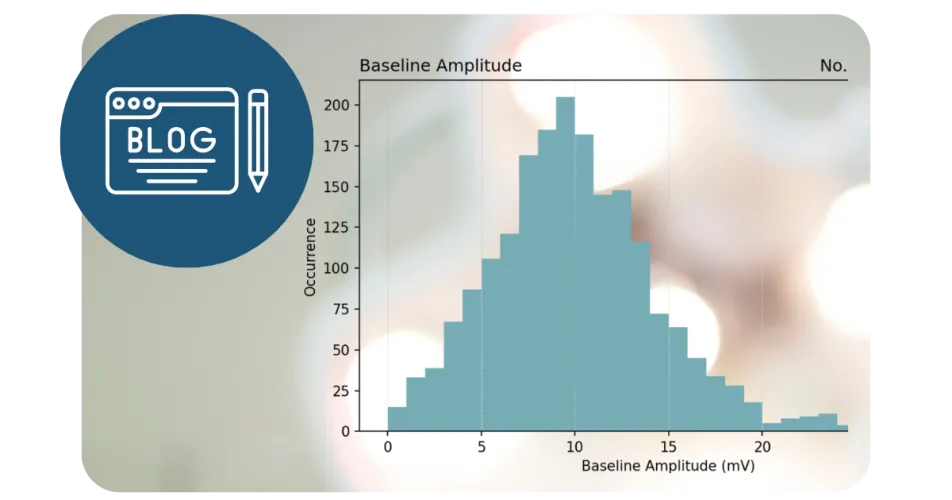
For years, the phrase “Strive for five” has been a cornerstone of training for quantitative train-of-four (TOF) monitoring with TetraGraph®. It remains a simple and effective guideline for achieving proper sensor placement and signal quality.
The recommendation is based on both clinical experience and science: a baseline EMG signal strength (amplitude) of at least 5 mV provides a reliable foundation for neuromuscular monitoring.
As we have gained experience from thousands of clinical cases and continued to refine the Next-generation TetraGraph platform, our guidance has evolved:
“Strive for 5, and 8 is great.”
Higher baseline signal strength (amplitude) provides greater robustness throughout the case. However, an important point is often overlooked:
A baseline amplitude below 5 mV does not necessarily mean inaccurate monitoring.
Next-generation TetraGraph is specifically designed to compensate for lower signal amplitudes and continue to provide reliable assessment of neuromuscular block.
Why Signal Strength Matters in Neuromuscular Monitoring
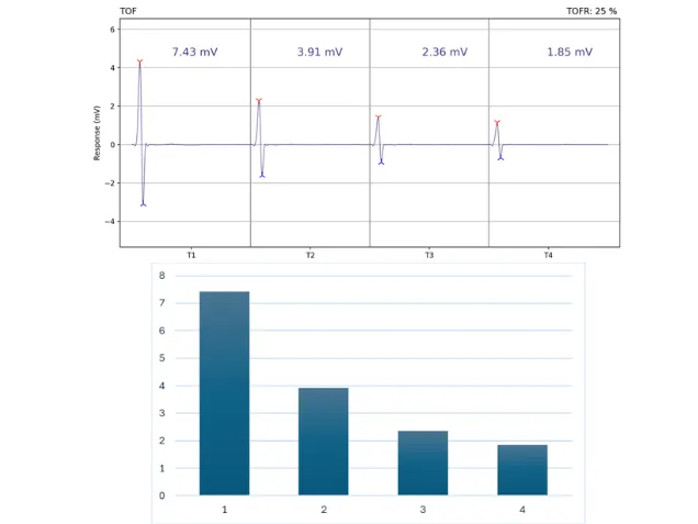
EMG-based monitoring measures the compound muscle action potential (CMAP) amplitude (height), providing a direct and quantitative assessment of neuromuscular function.
TetraGraph measures the initial signal from the nerve – the muscle action potential. Depicted is an example of a shallow depth of block (defined as TOF Ratio 10%-40%), where the patient’s EMG response is TOF Ratio=25% (1.85 mV/7.43 mV=25%).
The amplitude (peak-to-trough measurement) of the first waveform, T1 (expressed in millivolts) when monitoring is started before NMBA administration (pre-paralytic) is measured and displayed by the TetraGraph monitor as the baseline amplitude. This baseline or “initial signal strength” determines how much measurement bandwidth is available throughout the procedure. Higher signals (amplitudes) are generally more resilient to factors such as:
- Electrical noise
- Patient repositioning
- Sensor movement
- Changes in skin contact or resistance
As a result, stronger baseline signals (8 mV and above) typically provide:
- Greater signal stability throughout the case
- Reduced sensitivity to noise
- Lower risk of signal loss
- Improved signal detection during deep block (better detection of post-tetanic counts, PTC) and early recovery (earlier detection of transition from TOF Count=3 to TOF Ratio)
Therefore, optimizing signal strength at the start of monitoring remains an important best practice.
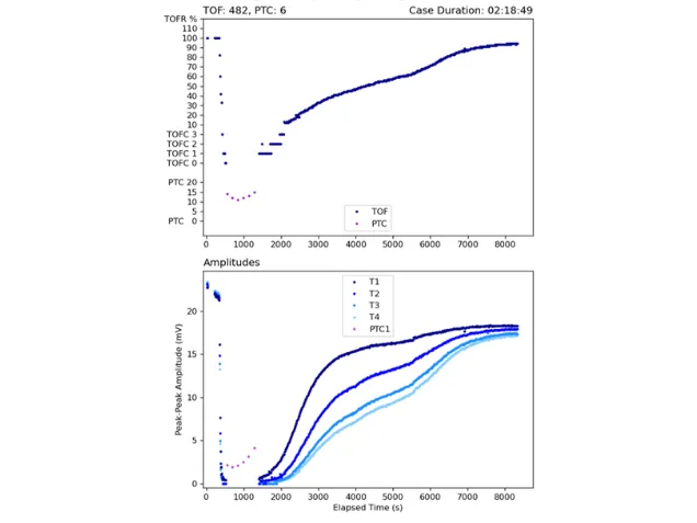
An example of a pediatric case with the trend view (upper graph) from case start (0) to finish (8000 s). This case demonstrates spontaneous recovery and high sensitivity from deep block and early recovery to acceptable recovery at TOFR >90%. The high amplitudes of the 4 individual twitches (T1-T4, visualized in the lower graph) ensure high sensitivity, tracking recovery with high precision.
What We See in Clinical Practice
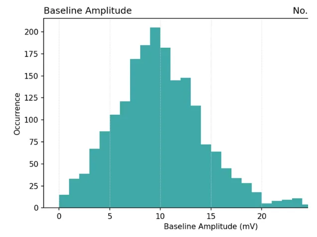
An analysis of 1,924 successful TetraGraph cases spanning from induction through recovery showed:
- Mean baseline signal strength: 9.99 mV
- Median baseline signal strength: 9.60 mV
These findings demonstrate that most users routinely achieve strong baseline signals, reflecting effective sensor placement and monitoring technique.
Further analysis revealed:
- 12% of cases started with a baseline amplitude below 5 mV
- 25% of cases started below 7 mV
- 34% of cases started below 8 mV
While most cases begin with signal strengths above 5 mV, lower baseline values are not uncommon in everyday clinical practice.
What Happens with Low Signal Strength?
A lower baseline signal from the start of the case primarily affects sensitivity, not accuracy.
When signal strength is low, the monitor has a smaller effective measurement window. This may reduce its ability to detect very small responses during:
- Deep neuromuscular block
- Early recovery, in the transition from TOF Count of 3 to TOF Ratio values.
In some cases, particularly after administration of sugammadex, the monitor may appear to transition rapidly from deep block directly to measurable TOF ratios because the earliest recovery responses are too small relative to the available signal window.
Importantly, this phenomenon affects only the earliest stages of recovery. Assessment of recovery remains reliable.
Even when baseline signal strength is lower than ideal:
- TOF Ratio measurements remain accurate
- Recovery to TOFR ≥0.90 is correctly identified
- Clinical decision-making at extubation remains reliable
This distinction is critical. A lower signal strength may reduce the monitor's ability to detect very small responses, but it does not compromise the accuracy of clinically relevant recovery measurements.
What Happens if Signal Strength Decreases During Surgery?
Signal strength (amplitude) can, and often does, decrease over time, and several factors prevent return of T1 to the initial control value. Acceleromyography (AMG) is particularly prone to baseline drift, and the final recovery T1 amplitude is often substantially lower than the initial control amplitude, unlike MMG and EMG, which show less drift on recovery.
Signal strength decrease is due to factors such as:
- Sensor loosening caused by patient repositioning
- Changes in electrode-skin contact
- Skin temperature changes
- Electrode impedance shifts
- Changes in muscle preload or position
- Changes in resting muscle tension
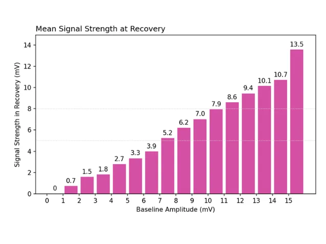
We have analyzed initial signal strength compared to the signal strength at the end of a case and documented that a high initial signal strength provides a higher likelihood of having a signal strength >5 mV at the end of a case.
- Initial signal 5–6 mV: mean recovery signal ≈ 3-4 mV
- Initial signal 7–8 mV: mean recovery signal ≈ 5-6 mV
When this occurs, the monitor may operate with reduced sensitivity and a smaller measurement window than from the start of the case (but without affecting precision). As with low baseline signals, this primarily affects the detection of very small responses during deep block and early recovery.
However, next-generation TetraGraph continues to provide accurate recovery assessment and correct TOF Ratios, including reliable identification of acceptable recovery at TOFR ≥0.90.
Impact on Clinical Workflow
Our data analysis also suggests that cases with lower signal strength are somewhat more likely to be ended before full recovery is documented. This is likely related to reduced sensitivity during early recovery and the assumption that "the monitor is stuck."
For this reason, achieving a strong baseline signal helps maximize visibility throughout the entire recovery process and provides greater confidence when following recovery trends over time.
The key point is that recovery accuracy remains intact even when signal strength decreases toward the end of a case: the clinician can confidently rely on TOF Ratio readings. It’s always recommended to continue to measure neuromuscular function until acceptable recovery of TOFR >90% is reached and to trust the monitor to display acceptable recovery accurately.
Practical Recommendations to Optimize Performance
- Aim for a baseline signal strength of 8 mV or higher whenever possible.
- A baseline of 5 mV or higher is generally sufficient for reliable monitoring.
- If baseline signal strength is below 5 mV, consider improving electrode placement and skin contact.
- Be aware that low signal strength may reduce sensitivity during deep block and early recovery but most importantly: Low signal strength does not mean inaccurate recovery monitoring.
While higher baseline amplitudes provide greater robustness and sensitivity, next-generation TetraGraph is designed to continue delivering accurate and clinically reliable TOF ratio measurements, including confirmation of recovery to TOFR ≥ 0.90.
Me and my colleagues are happy to guide you and answer any questions you might have.
Linnea Troeng, Sr Global Manager, Products and Business Development at Senzime